Cardiac Motion Segmentation
Project Summary
Current treatment methods for cardiac arrhythmia include medication, pacemakers, cardioversion, surgery, and fluoroscopy-guided radiofrequency ablation. In particular, radiofrequency ablation is recommended in many cases, due to its less invasive nature. However, there are still risks associated with it, such as injury to the heart, including perforation through the muscle or damage to one of the valves within the heart, exposure to radiation, which increases risk of cancer and genetic defects. MRI-guided radiofrequency ablation of the heart offers several advantages over current techniques, such as elimination of radiation exposure.

Currently most of the visualization for such procedures is done by either 2-D fluoroscopy or manual segmentation from 3-D CT models. On one hand, since fluoroscopy gives a 2-D projection of the 3-D object, it is difficult to accurately determine a 3-D location. On the other hand, manual segmentation is time consuming and suffers from reliability and consistency issues. Clearly, the improvement of 3-D visualization during the procedure could potentially help the physician locate the target more efficiently as well as eliminate catheter-induced injury to the heart.

In this project, we are developing methods for automatically building and updating a 3-D model of the heart using real time MR images. We first build a high-resolution 3-D model of the heart using MR and CT images acquired prior to the procedure. 
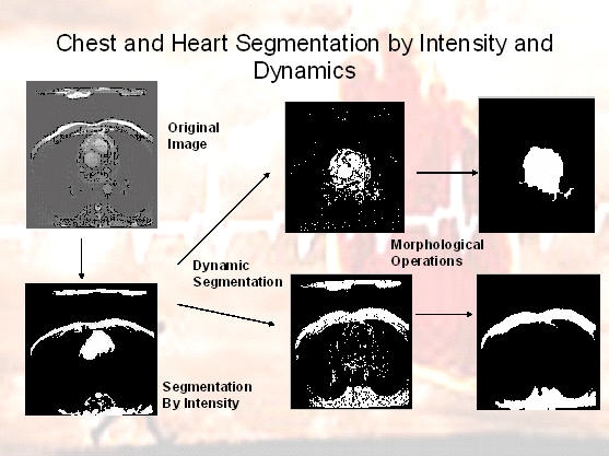
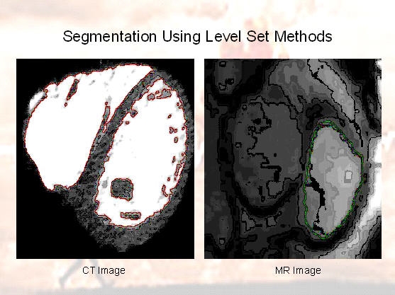
For accurate heart segmentation, two different methods have been presented: an fast algebraic method that does not impose hard spatial coherence hence smoothness, and a level set method, which is iterative but imposing smoothness. Given MRI data sequences, the heart and chest are separated from the background using the former method. In addition, we use dymanic textures to segment regions with different motions. The latter is used to segment different regions from the heart as well as to obtain smoother heart segmentation. We then build a 3-D high-resolution dynamic model that is updated with 2-D low-resolution images acquired in real time. In brief, the problems we try to solve are:
  1. Algebraic Heart/Chest/Background Segmentation
  2. Level Set based Epicardial Segmentation
  3. Segmentation Within The Heart
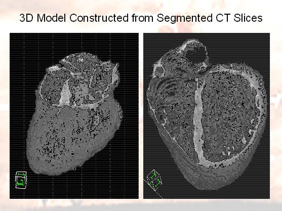
  4. Building The 3-D Model
Algebraic Heart/Chest/Background Segmentation
An MR axial image consists of three components: the heart, the chest and the background (which is predominantly noise). Given a volume of cardiac MR images, we consider the problem of segmenting the heart based on intensity and dynamics. In [1], we employ an algebraic technique for intensity-based segmentation called Polysegment to separate the heart and chest from the background. As the heart and the chest exhibit different dynamics, we model the image temporal evolution as the observations of two different linear dynamical systems. In other words, the remaining groups modeled as dynamic textures, and hence the trajectories of the heart and chest intensities lie in different subspaces. We use a method called Spatial Generalized Principle Component Analysis (Spatial GPCA) for clustering data points lying in multiple subspaces. Figure 1 shows the results of our segmentation algorithm.


Figure 1: Heart/Chest/Background Segmentation.

Level Set based Epicardial Segmentation
Level set methods are widely used in cardiac image segmentation because not only do they result in coherent regions with smooth boundaries, but also it is easy to incorporate prior information. Such methods perform point-wise or local operations on the image to find candidate points on the edge, and introduce the edge as the connection of the points. They start with a closed curve, minimize some energy function, and deform accordingly till they reach their optimal state.

In [2], we propose an alternative method for epicardial segmentation in dynamic MR sequences based on levet sets using priors on shape, intensity, and dynamics. The prior models are built from a training set of manually segmented sequences. The dynamics of the heart and background intensities are modeled with linear autoregressive models whose parameters are learnt from training data and then used as priors for segmentation. The segmented training shapes are registered with respect to a common reference. Each registered shape is represented with a signed distance function, and a statistical shape model is learnt using PCA on these functions. Segmentation is achieved by minimizing a spatial temporal generalization of the Mumford-Shah energy functional in a level set framework. Figure 2 shows the results of our algorithm.


Figure 2: Epicardial segmentation in different slices. Green contour: initialization. Red contour: final segmentation.

Segmentation within The Heart
We segment different regions of the heart in MR images using level sets. As mentioned before the main advantage of this method in this particular application is that it results in closed coherent areas with smooth boundaries, whereas in other methods, the edge is not guaranteed to be continuous or closed. Furthermore, using level set methods we can easily take care of breaking and merging of the curves with time, which is frequently seen in 2D MR images. Figure 3 shows the results of our algorithm.


Figure 3: Segmentation within the heart using level sets.

Building the 3-D model
We have volumized our segmentation results on a CT volume to get a 3D model of the heart with the blood taken out, and the background omitted.


Figure 4: 3-D Heart model.

Publications
[1]
A. Ravichandran, R. Vidal, and H. Halperin
IEEE International Symposium on Biomedical Imaging, pages 634-637, April 2006.
[2]
A. Ghoreyshi and R. Vidal.
IEEE International Symposium on Biomedical Imaging, pages 860-863, April 2007.