Orientation extraction and tracing of tubular structures in medical images are important quantitative tools for
developing models of the heart both at the histological and cytological levels. For instance, the configuration
of the myocardial fibers and the Purkinje network are crucial for modeling the mechanical and electrical properties
of the heart and understanding structural changes with myocardial infarction or arrhythmias. Likewise, the cytological
mechanical properties are related to the spatial orientation of the cytoskeletal filaments. In addition, detection
and enhancement of human vasculature is important for giving insight before interventional procedures.
The problems of detecting orientation and tracing tubular structures are closely related to a primal problem in image
processing and computer vision: edge detection. Existing approaches to solve those problems are based on the image Hessian H(x)
or the structure tensor S(x) at the point of interest x. Consider a grayscale image I:&upsih &rarr &real, &upsih being the image
domain. While H(x) possesses 2
nd order derivative information, i.e. H(x) = [I
xx I
xy; I
yx
I
yy], the structure tensor S(x) has 1
st order derivative information, S(x) = [I
x2
I
xI
y; I
yI
x I
y2].
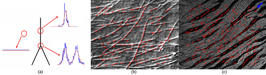
The eigendecomposition of the matrix H (or S) gives the orientation information: the eigenvector associated with the leading
eigenvalue is in the direction of the maximum gradient (perpendicular to the structure) and the remaining eigenvector shows the
tangential direction (along the structure). However, in case of intersections or bifurcations, it has the drawback of
"averaging" the orientation information built in the true structure. In our problem we have to deal with not only the image noise
but also the complexity such as intersection of fibers and bifurcation of the structure, which make the aforementioned approaches
useless.
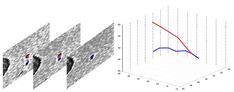
In this project, we consider the problem of extracting spatial orientation and tracing 2-D and 3-D tubular structures in medical
images.The problems we try to solve are:
- Local orientation extraction in myofiber arrays.
- Tracing tubular structures such as fibers, vessels, Purkinje network, etc. in medical images.