Diffusion Magnetic Resonance Imaging
Project Summary
Diffusion Magnetic Resonance Imaging (DMRI) is a medical imaging technique that is used to estimate the anatomical network of neuronal fibers in the brain, in vivo, by measuring and exploiting the constrained diffusion properties of water molecules in the presence of bundles of neurons. Water molecules will diffuse more readily along fibrous bundles (think of fiber optics cables), then in directions against them. Therefore by measuring the relative rates of water diffusion along different spatial directions, we can estimate the orientations of fibers in the brain. In particular, one important type of DMRI technique that we will analyze is high angular resolution diffusion imaging (HARDI) which measures water diffusion with an increased level of angular resolution in order to better estimate the probability of fiber orientation, known as the Orientation Distribution Function (ODF). HARDI is an advancement over the clinically popular Diffusion Tensor Imaging (DTI) which requires less angular measurements because of a Gaussian assumption which restricts the number of fiber orientations that can be estimated in each voxel. More accurate estimates of ODFs at the voxel level using HARDI lead to more accurate reconstructions of fiber networks. For instance, the extraction of neuronal fibers from HARDI can help understand brain anatomical and functional connectivity in the corpus callosum, cingulum, thalamic radiations, optical nerves, etc. DMRI has been vital in the understanding of brain development and neurological diseases such as multiple sclerosis, amyotrophic lateral sclerosis, stroke, Alzheimer's disease, schizophrenia, autism, and reading disability. To make DMRI beneficial in both diagnosis and clinical applications, it is of fundamental importance to develop computational and mathematical algorithms for analyzing this complex DMRI data. In this research area, we aim to develop methods for processing and analyzing HARDI data with an ultimate goal of applying these computational tools for robust disease classification and characterization.

Current research areas include:

  1. SPARSE HARDI RECONSTRUCTION. To develop advanced algorithms for the sparse representation and reconstruction of HARDI signals with the goals of speeding up HARDI acquisition and compact data compression.
  2. ODF ESTIMATION. To develop advanced algorithms for computing accurate fields of Orientation Distribution Functions (ODFs) from HARDI data.
  3. HARDI FEATURE EXTRACTION. To develop methods for extracting features from high-dimensional HARDI data that can be exploited for ODF clustering, fiber segmentation, HARDI registration and disease classification.
  4. HARDI REGISTRATION. To develop advanced algorithms for the registration of HARDI brain volumes to preserve fiber orientation information.
  5. DISEASE CLASSIFICIATION. To develop advanced classification techniques using novel HARDI feature representations to robustly classify and characterize neurological disease.
ODF Estimation
To estimate the orentation of fibers in each voxel of a HARDI brain dataset, orientation distribution functions (ODFs) are estimated from the HARDI signal. ODFs are probability distributions that live on the unit sphere and therefore must be nonnegative. Our work on ODF estimation enforces nonnegativity not only computationally on discrete grid points [1] but theoretically on the entire continuous spherical domain [2,3]. Enforcing nonnegativity is important for estimating accurate ODFs used for downstream processes of tractography, registration, segmentation and disease classification. In our most recent work, we consider that to enforce nonnegativity everywhere on a continuous domain, this equates to an infinite number of optimiztion contraints which is intractable. But even with a large number of constraints, many will be redudant. In fact, if we knew the minimum of the function, a priori, only one nonnegativity constraint at this point would suffice to enforce nonnegativiety everywhere. The work of [3] constructs an algorithm to select the optimal constraint needed to enforce ODF nonnegativity everywhere.

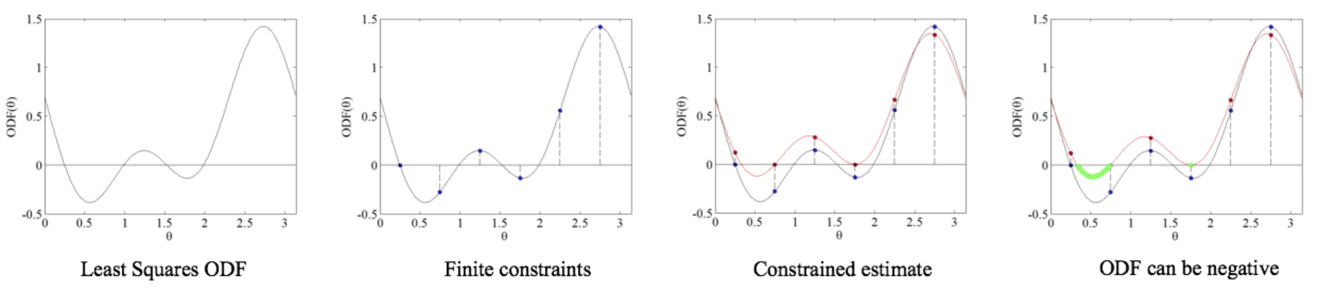
Figure 1: Illustration of nonnegativity enforcement on discrete set of points may leave continous functions negative. This calls for algorithms to enforce nonnegativity on the coninuous domain.
HARDI Feature Extraction
Reducing the amount of information stored in diffusion MRI (dMRI) data to a set of meaningful and representative scalar values is a goal of much interest in medical imaging. Such features can have far reaching applications in segmentation, registration, and statistical characterization of regions of interest in the brain. In fact, extracting scalar features from dMRI data has become an integral part of group/longitudinal studies of changes in brain connectivity related to development, neurodegeneration, or disease. Common features used in DTI are FA and MD and GFA for HARDI. However, existing features discard much of the information inherent in dMRI and embody several theoretical shortcomings and physical ambiguities. In this work [4] we propose a new framework for extracting a large set of rotation invariant features from the spherical harmonic (SH) representation of HARDI signals and ODFs. The advantage of this framework is its generality. In fact, we derive a family of rotation invariant features that can be extracted from any spherical function written in an SH basis. Numerous HARDI reconstruction methods such as Spherical Deconvolution (SD), Diffusion Orientation Transform (DOT), Spherical Polar Fourier Imaging (SPFI), Bessel Fourier Orientation Reconstruction (BFOR) model spherical functions like the Ensemble Average Propagator (EAP), Fiber Orientation Distribution (FOD), and Apparent Diffusion Coefficient (ADC) using an SH basis. Our framework can be applied to any of these spherical functions to extract a new set of scalar values. In addition, any continuous function of these scalar values can be used to generate additional features that can be significant for a specific experiment or application.

Figure 2: ISBI 2013 HARDI Phantom. First Row: The left image is the ground truth fiber segmentation of a slice of the phantom dataset, where we've identified an intricate region of crossing fibers. The right image is a count of the number of fibers that cross in a given voxel, ranging from 0 to 3. Second Row: GFA and eigenvalue variance of the phantom slice. We notice here the striking similarity between the plot of crossing fibers and the eigenvalue variance whereas the GFA is unable to reveal this information. Third/Fourth Row: Close up of the ROI with ODFs.
Publications
[1]
A. Goh, C. Lenglet, P.M. Thompson, and R. Vidal.
In Medical Image Computing and Computer Assisted Intervention, pp. 877-885, 5761, 2009.
[2]
E. Schwab, B. Afsari, and R. Vidal.
In Medical Image Computing and Computer Assisted Intervention, pp. 322-330, 7511, 2012.
[3]
S. Wolfers, E. Schwab, and R. Vidal.
In IEEE International Symposium on Biomedical Imaging, 2014.
[4]
E. Schwab, H. E. Cetingul, B. Afsari, M. A. Yassa, and R. Vidal.
In Information Processing in Medical Imaging, 2013.